


PROF. DR. HEDİYE DAĞDEVİREN
Gynecology, Obstetrics and IVF Specialist
Free Online Interview
Cant you have a child? Have you had unsuccesful attempts?

Gynecology, Obstetrics and IVF Specialist
Prof. Dr. Hediye Dağdeviren
Who?
She is a gynecologist and obstetrician born in 1980. She graduated from Dicle University in 2005. After working at Bakırköy Sadi Konuk Hospital and Kanuni Sultan Süleyman Training and Research Hospital, she is currently serving at VM Medical Park Hospital in Florya.

IVF Treatment Stages


In the process of stimulating the eggs, which is the first step in IVF treatment, the aim is to enlarge the egg cells in the ovary.
Egg collection is the first stage of the IVF process.
Perhaps the most important stage of IVF treatment is the process of combining the sperm and egg.
Embryo transfer is the process of placing embryos obtained outside the body into the uterus.
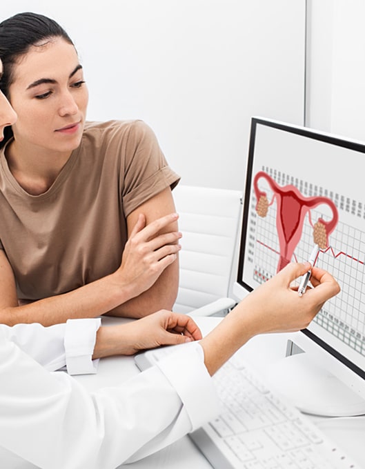
INFERTILITY
Infertility is defined as the inability to conceive within 1 year despite the regular and unprotected intercourse of the couple.
Free Online Interview
Week by Week Pregnancy
All pregnancy weeks together (9 Months 10 Days - 40 Week Summary)


Prof. Dr.
Hediye Dağdeviren
Hediye Dağdeviren
Gynecology, Obstetrics and IVF Specialist
©2022 Prof. Dr. Hediye Dağdeviren All Rights Reserved